| seg |
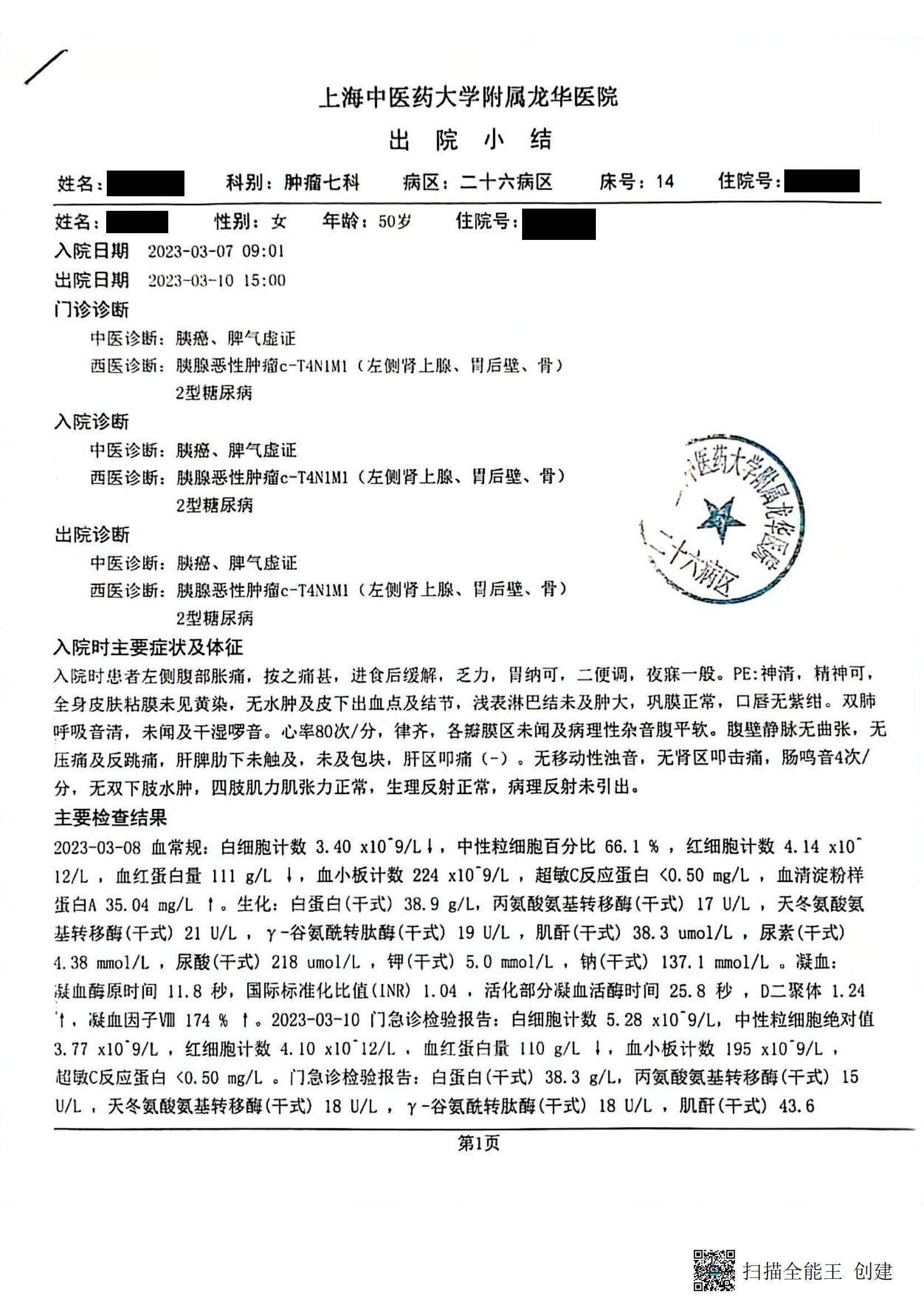
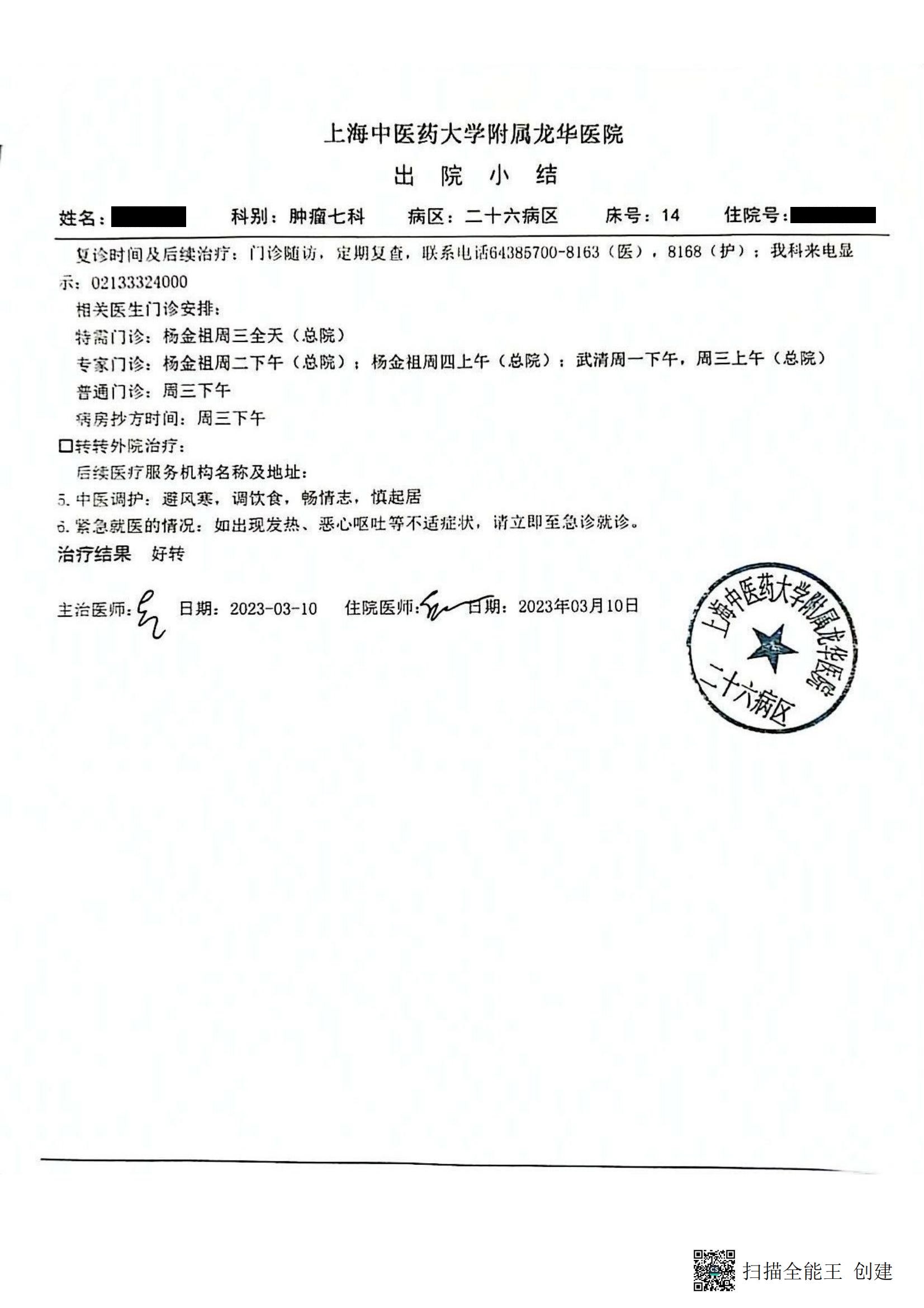
| 上海中医药大学附属龙华医院出院小结 |
| Discharge Summary of Longhua Hospital Affiliated to Shanghai University of Traditional Chinese Medicine |
| 姓名:*** 科别:肿瘤七科 病区:二十六病区 床号:14住院号:0823817 |
| Name: *** Department: Oncology the 7th Department Ward No.: 26 Bed No.: 14 Hospital No.: 0823817 |
| 更诊时间及后续治疗:门诊随访,定期复查,联系电话64385700-8163(医),8168 (护);我科来电显示:02133324000 |
| Time in service of hospital and follow-up treatment: outpatient follow-up, and regular review, Contact Number 64385700-8163 (medical), 8168 (nursing); Tel. Number of our department: 02133324000 |
| 相关医生门诊安排: |
| Outpatient Arrangements of Related MD : |
| 特需门诊:杨金祖 周三全天(总院) |
| VIP Clinic: Jinzu Yang all day on Wednesday (General Hospital) |
| 专家门诊:杨金祖 周二下午(总院);杨金祖 周四上午(总院):武清 周一下午,周三上午(总院) |
| Specialist clinic: Yang Jinzu Tuesday afternoon (General Hospital); Yang Jinzu Thursday morning (General Hospital): Wuqing Monday afternoon, Wednesday morning (General Hospital) |
| 普通门诊:周三下午 |
| General clinic: Wednesday afternoon |
| 病房抄方时间:周三下午 |
| Time for copying prescriptions in the ward: Wednesday afternoon |
| 转外院治疗: |
| Transfer to another hospital for treatment: |
| 后续医疗服务机构名称及地址: |
| Name and address of follow-up medical service institution: |
| 中医调护:避风寒.调饮食,畅情志,慎起居 |
| Traditional Chinese medicine care: avoid wind and cold, adjust diet, smooth emotions, and be cautious in daily life |
| 紧急就医的情况:如出现发热、恶心呕吐等不适症状,谙立即至急诊就诊。 |
| Emergency medical treatment: If symptoms such as fever, nausea, or vomiting occurs, go to the emergency department immediately. |
| 治疗结果 好转 |
| Treatment results: Improved |
| 主治医师: |
| Attending Physician: |
| 日期:2023-03-10 |
| Date: Mar. 10, 2023 |
| 住院医师: 日期:2023年03月10日 |
| Residency Physician: Date: Mar. 10, 2023 |
| 爨扫描全能王创建 |
|
| 上海中医药大学附属龙华医院出院小结 |
| Discharge Summary of Longhua Hospital Affiliated to Shanghai University of Traditional Chinese Medicine |
| 姓名:*** 科别:肿瘤七科 病区:二十六病区 床号:14住院号:0823817 |
| Name: *** Department: Oncology the 7th Department Ward No.:26 Bed No.: 14 Hospital No.: 0823817 |
| umol/L ,尿素(干式)5. 01 mmol/L ,尿酸(干式)307 umol/L,钾(干式)4. 1 mmol/L,钠(干式) 132.9 mmol/L . |
| umol/L, urea (dry) 5.01 mmol/L, uric acid (dry) 307 umol/L, potassium (dry) 4.1 mmol/L, sodium (dry) 132.9 mmol/L. |
| 特殊检查结果及主要会诊(注明日期与检查号) 无 |
| Special inspection results and major consultations (specify date and inspection number) : None |
| 病程与治疗结果(注明手术日期、手术名称、输血以及抢救情况) |
| Disease course and treatment results (indicate operation date, operation name, blood transfusion and rescue status) |
| 患者四月前因反复腹部隐痛不适,遂于2022. |
| The patient had abdominal pain and discomfort repeatly in four months ago, and then |
| 9月至复旦大学附属肿瘤医院就诊,2022.09. 15肿瘤标志物:CA199:7459U/ml, CA125:79. 2U/ml, CA153:4. 94U/ml, CA724:2. 32U/ml, CA50: >500IU/ml, CA242: > 200U/ml. |
| visited Fudan University Shanghai Cancer Center on Sept. 15 2022. Tumor markers: CA199: 7459U/ml, CA125: 79. 2U/ml, CA153: 4. 94U/ml, CA724: 2. 32U/ml, CA50: > 500IU/ml, CA242: > 200U/ml. |
| AFP:2.43ng/ml, CEA: 165ng/ml. |
| AFP: 2.43ng/ml, CEA: 165ng/ml. |
| 2022-09-15 查【PET-CT】报告示:1.胰体部软组织肿块影,FDG 代谢增高,考虑为MT可能大,侵犯左侧肾上腺及胃后壁。 |
| 2022-09-15 [PET-CT] report showed: 1. Soft tissue mass shadows in the body of the pancreas, increased FDG metabolism, it is considered that the MT possibiity is high, invading the left adrenal gland and the back wall of the stomach. |
| 2022. 09. 20行超声引导下胰体尾肿瘤穿刺,2022- 09-21穿刺细胞病理诊断示:见腺癌细胞,遂于肿瘤医院2022-09-22至2023-01-12行AG方案化疗(紫杉醇 170mg dl+吉西他滨1.4g dl) 4程.2023-01-31于复旦大学附属肿瘤医院[PET-CT]示:1.胰腺癌化疗后,胰体MT较前相仿,仍FDG代谢异常增高,侵犯左侧肾上腺及胃后壁;新见胸骨、右侧股骨上段转移;新见两侧锁骨上淋巴结转移可能。 |
| 2022.09.20 Ultrasound-guided pancreatic tail tumor puncture, 2022-09-21 Puncture cytological diagnosis showed: adenocarcinoma cells were seen, and AG chemotherapy was performed in the Cancer Hospital from Sept. 22, 2022 to Jan. 12, 2023 (Paclitaxel 170mg dl + Gemcitabine 1.4g dl) 4 courses. On Jan. 31,2023 at Fudan University Cancer Hospital, the [PET-CT] showed: 1. After pancreatic cancer chemotherapy, pancreatic MT was similar as before, FDG metabolism was still abnormally increased, Invading the left adrenal gland and the posterior gastric wall; new metastasis to the sternum and right upper femur; new possible metastasis to supraclavicular lymph nodes on both sides. |
| 2023-02-09、2023-03-02我科行AG方案C5治疗:AG化疗方案(白蛋白紫杉醇200mg +吉西他滨L 4g ivgtt dl),此次入院后完善相关检查,患者病理诊断明确,ECOG评分合格,心 电图及生化检查合格,排除相关禁忌,经主任同意,2023-03-09行腹腔干+肠系膜上动脉化疗术(白蛋白紫杉醇100mg+吉西他滨0.4g IA),并子枢星止呕,爱丽安护胃,美能护肝。 |
| on Feb. 9, 2023 and Mar. 2, 2023, Our department performed C5 treatment of AG program: AG chemotherapy program (nab-paclitaxel 200mg + gemcitabine L 4g ivgtt dl), relevant examinations were completed after admission, the patient’s pathological diagnosis was clear, and the ECOG score was qualified, the electrocardiogram and biochemical examination were qualified, and the relevant contraindications were excluded. With the consent of the director, celiac trunk + superior mesenteric artery chemotherapy (nab-paclitaxel 100mg + gemcitabine 0.4g IA) was performed on March 09, 2023, with Granisetron Hydrochloride Tablets for relieving vomiting, Ilaprazole Enteric-coated Tablets for protecting the stomach, and SNMC for protecting liver. |
| 余治疗于门冬胰岛素30早12u-晚 11u皮下。 |
| Insulin aspart 30 was given with 12u in the morning and 11u in the afternoon as treatment by subcutaneous injection |
| 查患者舌淡红,苔薄白,脉细,证属“胰癌脾气虚证”,子通关藤、康艾清热解毒抗肿痛,正得康胶囊扶正抗肿瘤,配合耳针益气扶正。 |
| The patient had a pale red tongue, thin white fur, and thready pulse. The syndrome belongs to “pancreatic cancer with spleen deficiency syndrome”. Medicines of Marsdeniae Tenacissimae Caulis,Kang‘ai Zhusheye,Zhengdekang capsule and acupunctur threapy are given for cancer treatment. |
| 经治疗,患者症情缓解,经上级医师同意,准予出院。 |
| After that, the patient’s symptoms were relieved, and the patient was allowed to leave the hospital with the consent of the superior physician. |
| 合并症 |
| Complications |
| 无 |
| None |
| 出院时情况(症状与体征) 出院时患者左侧腹部胀痛较前缓解,无明显不适,乏力较前缓解,胃纳可,二便调,夜寐一般“查体:神清,精神可,全身皮肤粘膜未见黄染,无水肿及皮下出血点及结节,浅表淋巴结未及肿大,巩膜正常,口唇无紫绀。 |
| Health status at discharge (symptoms and signs) When discharged from the hospital, the left abdomen pain of the patient was relieved without obvious discomfort, fatigue was relieved, appetite was acceptable, night soil and urine were normal, and night sleep was normal. No jaundice was found on the skin or mucous membranes of the whole body, no edema, no subcutaneous bleeding points or nodules, superficial lymph nodes were not enlarged, the sclera was normal, and the lips were not cyanotic. |
| 双肺呼吸音清,未闻及干湿啰音。 |
| Breath sounds in both lungs were clear. Dry or wet rales were not heard. |
| 心率76次/分,律齐,各瓣膜区未闻及病理性杂音腹平软。 |
| The heart rate was 76 beats/min and regular, No pathological murmurs were heard in each valve area, and the abdomen was flat and soft. |
| 腹壁睁脉无曲张,无压痛及反跳痛,肝脾肋下未触及,未及包块,肝区叩痛(-)。 |
| No varicose veins in the abdominal wall, no tenderness or rebound pain, no palpation of the liver and spleen under the ribs, no mass, There is pain on percussion in the liver area (-). |
| 无移动性浊音,无肾区叩击痛,肠鸣音4次/分,无双下肢水肿,四肢肌力肌张力正常,生理反射正常,病理反射未引出。 |
| No shifting dullness, no percussion pain in the kidney area, bowel sounds 4 times/min, no lower limb edema, normal muscle strength and muscle tone in the four limbs, normal physiological reflexes, and no pathological reflexes. |
| 出院后用药及建议 |
| Medication and advice after discharge |
| .出院带药:得康胶囊*1盒 每日三次,每次4粒 口服 |
| Discharge medicine: Dekang Capsule*1 box, three times a day, 4 capsules each time orally |
| .自备药:门冬胰岛索30 早12u-晚11u皮下。 |
| Self-prepared medicine: insulin aspart 30 12u-in the morning 11u at night by subcutaneous injection. |
| .药物潜在副作用:详见说明书 |
| . Potential side effects of the medicine: see the instructions for details |
| .后续医疗服务的安排:门诊随访,定期及查,如有不适及时就诊. |
| .Arrangement of follow-up medical services: outpatient follow-up, regular check-ups, and timely visit hospital if there is any discomfort. |
| 本院门诊随访 |
| Outpatient follow-up in our hospital |
| m扫描全能王创建 |
|
| / |
| / |
| 上海中医药大学附属龙华医院出院小结 |
| Discharge Summary of Longhua Hospital Affiliated to Shanghai University of Traditional Chinese Medicine |
| 姓名:*** 科别:肿瘤七科病区:二十六病区床号:14 住院号:0823817 |
| Name: *** Department: Tumor the 7th Department Ward No.: 26 Bed No.: 14 Hospital No.: 0823817 |
| 姓名:*** 性别:女年龄:50岁住院号:0823817 |
| Name: *** Gender: Female Age: 50 Admission No.: 0823817 |
| 入院日期 2023-03-07 09:01出院日期 2023-03-10 15:00门诊诊断 |
| Admission Date Mar. 7, 2023 09:01 Discharge Date Mar. 10, 2023 15:00 Outpatient Diagnosis |
| 中医诊断:胰癌、脾气虚证 |
| Traditional Chinese Medical Diagnosis: Pancreatic Cancer, Spleen Deficiency Syndrome |
| 西医诊断:胰腺恶性肿痛C-T4N1M1 (左侧肾上腺、胃后壁、骨)2型循尿病 |
| Western medicine diagnosis: pancreatic malignant swelling and pain C-T4N1M1 (left adrenal gland, back wall of stomach, bone) type II diabetes |
| 入院诊断 |
| Admission diagnosis |
| 中医诊断:胰癌、脾气虚证 |
| Traditional Chinese Medical Diagnosis: Pancreatic Cancer, Spleen Deficiency Syndrome |
| 西医诊断:腹腺恶性肿瘤C-T4N1M1 (左侧肾上腺、胃后壁、骨)2型糖尿病 |
| Western medicine diagnosis: abdominal gland malignant tumor C-T4N1M1 (left adrenal gland, back wall of stomach, bone) type II diabetes |
| 出院诊断 |
| Discharge diagnosis |
| 中医诊断:胰癌、脾气虚证 |
| Traditional Chinese Medical Diagnosis: Pancreatic Cancer, Spleen Deficiency Syndrome |
| 西医诊断:胰腺恶性肿瘤C-T4N1M1 (左侧肾上腺、胃后壁、骨)2型糖尿病 |
| Western medicine diagnosis: pancreatic malignant tumor C-T4N1M1 (left adrenal gland, back wall of stomach, bone) type 2 diabetes mellitus |
| 入院时主要症状及体征 |
| Main symptoms and signs on admission |
| 入院时患者左侧腹部胀痛,按之痛甚,进食后缓解,乏力,胃纳可,二便调,夜寐一股。 |
| When admitted to the hospital, the patient had distending pain in the left abdomen, which was very painful when pressed, and was relieved after having food. fatigue, normal appetite, bowels and urine open, and normal night sleep. |
| PE:神清,精神可,全身皮肤粘膜未见黄染,无水肿及皮下出血点及结节,浅表淋巴结未及肿大,巩膜正常,口唇无紫绀。 |
| PE: good consciousness, energetic, no jaundice in skin mucosa of the whole body, no edema, subcutaneous bleeding points or nodules, no superficial lymph node enlargement, normal sclera, and no cyanosis of lips. |
| 双肺呼吸音清,未闻及干湿啰音。 |
| Breath sounds in both lungs were clear. Dry or wet rales were not heard. |
| 心率80次/分,律齐,各瓣膜区未闻及病理性杂音腹平软。 |
| The heart rate was 80 beats/min and regular, no pathological murmurs were heard in each valve area, and the abdomen was flat and soft. |
| 腹壁静脉无曲张,无 压痛及反跳痛,肝脾肋下未触及,未及包块,肝区叩痛(-).无移动性浊音,无肾区叩击痛,肠鸣音4次/分,无双下肢水肿,四肢肌力肌张力正常,生理反射正常,病理反射未引出。 |
| No varicose veins in the abdominal wall, no tenderness or rebound pain, no palpation of the liver and spleen under the ribs, no mass, percussion pain in the liver area (-). No shifting dullness, no percussion pain in the kidney area, bowel sounds 4 times/min. No lower limbs edema, normal muscle strength and muscle tone of four limbs, normal physiological reflex, no pathological reflex elicited. |
| 主要检查结果 |
| Main inspection results |
| 2023-03-08血常规:白细胞计数3. |
| 2023-03-08 Blood routine: white blood cell count |
| 40 x109/L ,中性粒细胞百分比66. |
| 3.40 x10^9/L , neutrophil percentage 66. |
| 1 % ,红细胞计数4.14 xlO12/L ,血红蛋白量111 g/L ,血小板计数224 x109/L ,超敏C反应蛋白<0. 50 mg/L ,血清淀粉样蛋白A 35. 04 mg/L。 |
| 1%, red blood cell count 4.14 x10^12/L, hemoglobin 111 g/L, platelet count 224 x10^9/L, hypersensitive C-reactive protein <0. 50 mg/L, serum amyloid A 35. 04 mg/L. |
| 生化:白蛋白(干式)38.9 g/L,丙氨酸氨基转移酣(干式)17 U/L ,天冬氨酸氨基转移酶(干式)21 U/L . γ-谷氨酰转肽酶(干式)19 U/L ,肌酐(干式)38.3 umol/L ,尿素(干式) 4. 38 mmol/L , 尿酸(干式)218 umol/L .钾(干式)5. 0 mmol/L,钠(干式)137. 1 mmol/L。 |
| Biochemical: Albumin (dry) 38.9 G/L, Alanine Transaminotransferase (dry) 17 U/L, Aspartate Aminotransferase (dry) 21 U/L. Γ-glutamyl Transfer Peptidase (dry) 19 U/L, Creatinine (dry) 38.3 Umol/L, Urea (dry) 4.38 Mmol/L, Uric Acid (dry) 218 Umol/L, Potassium (dry) 5. 0 Mmol/L, Sodium (dry ) 137.1 Mmol/L. |
| 凝血: 凝血酶原时间11.8秒,国际标准化比值(INR) 1.04,活化部分凝血活酶时间25.8秒,D二聚体1.24,凝血因子VIII174 % t . |
| Coagulation: Prothrombin Time 11.8 Seconds, International Normalized Ratio (INR) 1.04, Activated Partial Thromboplastin Time 25.8 Seconds, D Dimer 1.24, Coagulation Factor VIII 174 % |
| 2023-03-10门急诊检验报告:白细胞计数5. |
| 2023-03-10 Outpatient emergency test report: white blood cell count 5. |
| 28 xI09/L,中性粒细胞绝对值 3.77 xlO9/L .红细胞计数4. |
| 28 xI0^9/L, Absolute Value Of Neutrophils 3.77 XlO^9/L. Red Blood Cell Count 4. |
| 10 x10l2/L ,血红蛋白量110 g/L I ,血小板计数195 x109/L . 超敏C反应蛋白<0. 50 mg/L 。 |
| 10 x10^l2/L, Hemoglobin 110 g/L I, Platelet Count 195 X10^9/L. Hypersensitive C-reactive Protein <0. 50 mg/L. |
| 门急诊检验报告:白蛋白(干式)38.3 g/L,丙氨酸氨基转移酶(干式)15 U/L ,天冬氨酸氨基转移酶(干式)18 U/L , Y-谷氨酰转肽酶(干式)18 U/L ,肌酐(干式)43.6 |
| Outpatient and emergency test report: Albumin (dry ) 38.3 G/L, Alanine Aminotransferase (dry) 15 U/L, Aspartate Aminotransferase (dry) 18 U/L, Y-glucose Amyltranspeptidase (dry) 18 U/L, Creatinine (dry ) 43.6 |
| 第1页 |
| page 1 |
| m扫描全能王创建 |
|
| 复旦大学附属肿瘤医院 核医学科 |
| Cancer Hospital Affiliated to Fudan University Department of Nuclear Medicine |
| PET |
| PET |
| 检查报告单 |
| Physical Examination Report |
| 影像号:11407881 |
| Image number: 11407881 |
| 姓名:*** 性别1女 |
| Name: *** Gender: Female |
| 检查方式核素 |
| Examination method: nuclide |
| 申请科室 |
| Department Application |
| 临床论断: |
| Clinical conclusion: |
| PET/CT 断层 |
| PET/CT tomography |
| 药物: FDG胰腺外科 |
| Medicine: FDG Pancreatic Surgery |
| 胰腺癌化疗后 |
| Pancreatic Cancer After Chemotherapy |
| 年龄50 岁 |
| Age: 50 |
| 检查部位:全身 |
| Inspection Area: whole body |
| 剂量 6.048mCi |
| Dosage 6.048mCi |
| 检查日期,2023-01-31 |
| Inspection date, Jan. 31, 2023 |
| 门诊号 310104197210274823 |
| Clinic No. 310104197210274823 |
| 床号 |
| Bed No. |
| 血糖:6. 2mmol/L |
| Blood sugar: 6. 2mmol/L |
| 检查项目:肿瘤全身断层显像 |
| Inspection items: Tumor whole body tomography |
| 给药方式:静脉注射 注射部位:右手腕 |
| Administration method: intravenous injection Injection part: right wrist |
| 检查排号:25 |
| Queue number for examination: 25 |
| — |
| — |
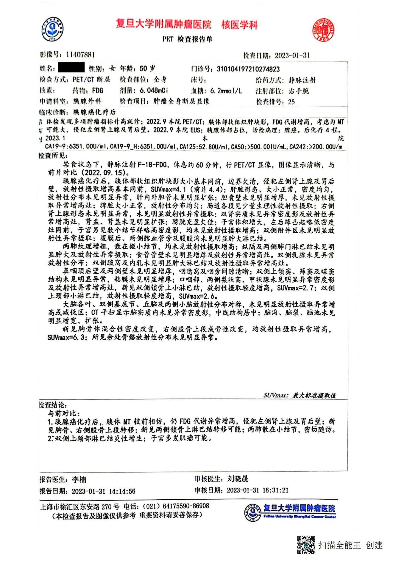
| 体检发现多项肿瘤指标升高就诊:2022. 9 本院PET/CT:肢体部软组织肿块影,FDG代谢增高,考再为MT可能大.侵犯左侧肾上腺及胃后壁.2022.9本院EUS:质腺体部占位,活检病理:腺癌.后化疗4程。 |
| A number of tumor indicators were found to be elevated in the physical examination and visit the hospital: In Sept., 2022. PET/CT in our hospital: soft tissue mass shadow in the limbs, FDG metabolism increased, high MT possibility was considered. Invasion of the left adrenal gland and the back wall of the stomach. Sept. 2022 EUS in our hospital: Gland space occupying, biopsy pathology: adenocarcinoma. Chemotherapy with 4 courses. |
| 2023. 1 |
| Jan. 2023 |
| 本院 |
| This hospital |
| CA19-9:6351. |
| CA19-9:6351. |
| OOU/mI, CA19-9_H:6351. 00U/mI, CA125:52. 80U/ml,CA50:>500. 00lU/mL, CA242:>200. 00U/m |
| OOU/mI, CA19-9_H:6351. 00U/mI, CA125:52. 80U/ml, CA50:>500. 00lU/mL, CA242:>200. 00U/m |
| 检杳所见: |
| Examination Result: |
| 禁食状态下.静脉注射F-18-FDG,体息约60分钟,行PET/CT显像,图像显示清晰,与前片对比(2022. 09.15). |
| In the fasting state. F-18-FDG was injected intravenously, and rested for about 60 minutes. PET/CT imaging was performed, and the image was clearly displayed, comparing with the previous film (Sept. 15, 2022). |
| 胰腺癌化疗后.肢体部软组织肿块影大小基本同前,边界欠清,侵犯左侧肾上腺及胃后壁,放射性摄取增高基本同前,SUVmax=4.1 (前片4.4): |
| After chemotherapy for pancreatic cancer, the size of the soft tissue mass in the extremities is basically the same as before, with unclear borders, invading the left adrenal gland and the back wall of the stomach, and the increase in radioactive uptake is basically the same as before, SUVmax=4.1 (4.4 in the previous film): |
| 肝脏形态、大小正常,密度均匀, 放射性分布未见明显异常.肝内外胆管皆未见明显犷张; |
| The shape and size of the liver were normal, the density was uniform, and there was no obvious abnormality in the distribution of radioactivity. There was no obvious swelling of the bile ducts inside and outside the liver; |
| 胆囊壁未见明显增厚,未见放射性摄取异常增高灶: |
| There was no obvious thickening of the gallbladder wall, and no abnormally increased radioactive uptake foci: |
| 脾脏大小正常,放射性分布均匀; |
| The size of the spleen is normal and the distribution of radioactivity is uniform; |
| 肠道各段见少量生理性放射性摄取: |
| A small amount of physiological radioactive uptake is seen in various segments of the intestinal tract: |
| 右侧肾上腺形态未见明显异常,未见明显放射性异常摄取; |
| There was no obvious abnormality in the shape of the right adrenal gland, and no obvious abnormal uptake of radioactivity; |
| 双肾实质未见异常密度影及放射性异常增高灶,肾孟、肾盏未见明显扩张; |
| There were no abnormal density shadows and abnormally increased radioactive foci in the renal parenchyma, and no obvious expansion of the renal calices and pelvis; |
| 膀胱充盈欠佳; |
| Poorly filling bladder; |
| 子客体积增大,左后缘凸起略低密度灶同前,子宫另见数个结节样略高密度影,均未见放射性摄取增高; |
| The volume of the uterus increases, the left posterior margin bulges slightly low density foci are the same as before, and several nodular with slightly high density shadows are seen in the uterus, all of which have no increased radiation uptake; |
| 双侧附件区未见明显放射性异常摄取: |
| No obvious abnormal radioactive uptake in the bilateral appendages: |
| 腹膜后、两侧髂血管旁及腹股沟未见明显肿大淋巴结。 |
| No obvious enlarged lymph nodes were found in retroperitoneum, beside bilateral iliac vessels or groin. |
| 两肺及理增粗,散在微小结节,均未见放射性摄取增高:纵隔及两侧肺门淋巴结未见明显肿大及放射性异常摄取:食管管壁未见明显增厚及放射性异常增高灶。 |
| The two lungs were thickened, with scattered tiny nodules, and no increased radioactive uptake was found: no obvious enlargement and abnormal radioactive uptake were found in the mediastinal and bilateral hilar lymph nodes; no obvious thickening of the esophageal wall and abnormally increased radioactive foci were found. |
| 双侧乳腺未见异常放射性分布:双侧腋窝及内乳未见明显肿大淋巴结及放射性摄取异常增高灶。 |
| No abnormal radiation distribution was found in bilateral breasts: no obvious enlarged lymph nodes or abnormally increased radioactive uptake foci were found in bilateral axillaries and inner breasts. |
| 鼻咽顶后壁及两侧壁未见明显增厚,咽隐窝及咽旁间隙清晰: |
| The posterior wall of the nasopharyngeal roof and both sides were not thickened, and the pharyngeal recesses and parapharyngeal space were clear: |
| 双侧上颌窦、筛窦及堞窦 结构未见明显异常,黏膜未见明显增厚: |
| There was no obvious abnormality in the structure of the bilateral maxillary sinus, ethmoid sinus, and castellation sinus, and no obvious thickening of the mucosa: |
| 口咽部、两侧梨状窝、甲状腺未见明显异常密度影及放射性异常增高灶,新见双侧锁骨上小淋巴结,放射性摄取轻度增高,SUVmax=2.7; |
| no obvious abnormal density shadow and abnormally increased radioactive focus at Oropharynx, bilateral pyriform sinuses or thyroid, new bilateral supraclavicular small lymph nodes appears with slightly increased radioactive uptake, SUVmax=2.7; |
| 双侧 上颈部小淋巴结,放射性摄取轻度增高,SUVmax=2. 6。 |
| The radioactive uptake was slightly increased in the small lymph nodes of the upper neck bilaterally, SUVmax=2.6. |
| 大脑各叶、双侧基底节、丘脑及两侧小脑放射性分布对称,未见明显放射性摄取异常增高或减低区;CT平扫显示脑实质内未见异常密度影,中线结狗居中:脑沟、脑裂、脑池未见明显增宽、扩张。 |
| The radioactivity distribution in each lobe of the brain, bilateral basal ganglia, thalamus and bilateral cerebellum is symmetrical, and there is no obvious abnormal increase or decrease of radioactivity uptake; CT plain scan showed that there was no abnormal density shadow in the brain parenchyma, and the midline structure was in the middle: the sulcus, fissure and cistern were not significantly widened or expanded. |
| 新见胸骨体混合性密度改变,右侧股骨上段成骨性改变,均放射性摄取异常增高, SUVmax=6. 3:所见余处骨骼放射性分布未见明显异常。 |
| The mixed density change of the sternum body and the osteogenic change of the right upper femur are newly seen, and the radioactive uptake is abnormally high, SUVmax=6 3: There is no obvious abnormality in the radioactive distribution of the remaining bones. |
| SUVmax;最大标准摄取值 检查结论「 |
| SUVmax; maximum standard intake value inspection conclusion |
| 与前对比: |
| Compared with before: |
| 1.肢腺癌化疗后,肢体MT较前相仿,仍FDG代谢异常增高,侵犯左侧肾上腺及胃后壁:新见胸骨、右侧股骨上段转移;新见两侧锁骨上淋巴结转秽可能:两肺散在小结节,密切随访。 |
| 1. After chemotherapy for pancreatic cancer, the limb MT was similar as before, but the FDG metabolism was still abnormally high, and the left adrenal gland and the posterior wall of the stomach were invaded: the sternum and the upper part of the right femur were newly found to metastasize; The newly seen bilateral supraclavicular lymph node metastasis is possible: scattered small nodules in both lungs, close follow-up is needed. |
| 2:双侧上颈部淋巴结炎性增生;子宫多发肌瘤可能。 |
| 2: Inflammatory hyperplasia of bilateral upper cervical lymph nodes; multiple fibroids of the uterus is possible. |
| 报告医生:李楠审核医生:刘晓晟 |
| Reporting doctor:Nan Li Reviewing doctor: Xiaosheng Liu |
| 报告日期:2023-01-31 14:14:56审核日期:2023-01-31 16:31:21 |
| Reporting Date: Jan. 31, 2023 14:14:56 Reviewing Date: Jan. 31, 2023 16:31:21 |
| 上海市徐汇区东安路270号电话:(021) 64175590-86908复旦大学附属肿病医院 |
| No. 270, Dong’an Road, Xuhui District, Shanghai Tel: (021) 64175590-86908 Cancer Hospital Affiliated to Fudan University |
| (本检查报告及图像仅供参考重要资料请妥善保存)tMwfKd C- |
| (This inspection report and images are for reference only, please keep the information properly) |
| m扫描全能王创建 |
|
| 复旦大学附属肿痛医院 核医学科 |
| Department of Nuclear Medicine, Cancer Hospital, Affiliated to Fudan University |
| PET 检查报告单 |
| PET inspection report |
| 影像号 11407881 |
| Image number 11407881 |
| 检查日期 2022/09/15 |
| Inspection date: Sept. 15, 2022 |
| 姓名:*** 性别:女 |
| Name: ***Gender: Female |
| 检查方式: PET/CT断层 |
| Inspection method: PET/CT tomography |
| 核素: 18F药物:FDG |
| Nuclide: 18F Medicine: FDG |
| 申请科室:胰腺外科 |
| Department Application: Pancreatic Surgery |
| 临床诊断:胰腺癌 |
| Clinical Diagnosis: Pancreatic Cancer |
| 年龄:49 8门诊号:310104197210274823 |
| Age: 49 Clinic No.: 310104197210274823 |
| 检查部位:全身 床号:给药方式:静脉注射 |
| Examination Area: Whole Body Bed No.: Administration Method: Intravenous Injection |
| 剂量: 5. 285mOI 血糖:10.0mmol/L 注射部位:右手背 |
| Dosage: 5. 285mOI Blood sugar: 10.0mmol/L Injection site: Back of right hand |
| 检查项目:肿瘤全身断层显像检查排号,46 |
| Inspection items: Tumor whole body tomography inspection Queue Number: 46 |
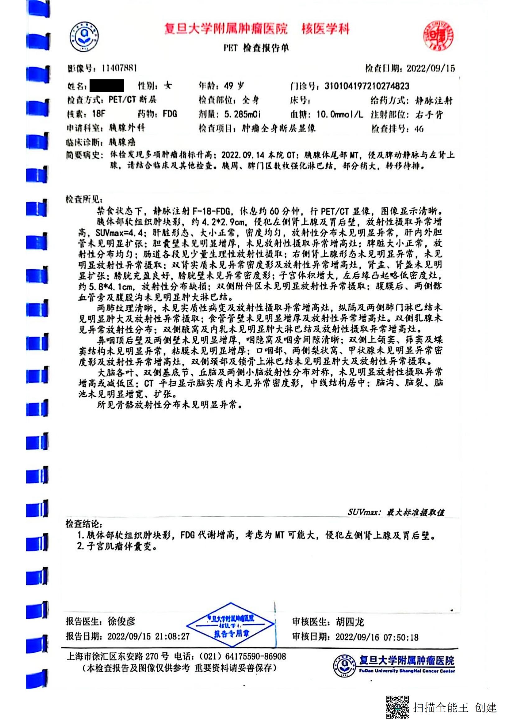
| 简要病史:体检发见多项肿瘤指标升高:2022.09. 14本院CT:胰腺体尾部MT,侵及脾动静脉与左肾上腺,请结合临床及其他其他检查。 |
| Brief medical history: Multiple tumor indicators increasing was found in physical examination: Sept. 14, 2022 CT in our hospital: MT of the pancreas body tail, invading the splenic arteriovenous and left adrenal gland, please combine with clinical and other examinations. |
| 胰周、脾门区数枚强化淋巴结,部分稍大,转修待排. |
| There were several enhanced lymph nodes in the peripancreatic and splenic hilum area, some of which were slightly larger. |
| 检查所见: |
| Examination Results: |
| 禁食状态下,静脉注射F-18-FDQ,休息约60分钟,行PET/CT显像,图像显示清晰. |
| In the fasting state, F-18-FDQ was injected intravenously, rested for about 60 minutes, and PET/CT imaging was performed, and the images were clearly displayed. |
| 胰体部软组织肿块影,约4.2*2.9cm.侵犯左侧肾上腺及胃后壁,放射性摄取异常增 高,SUVmax=4.4: |
| The soft tissue mass shadow in the body of the pancreas is about 4.2 * 2.9 cm. It invades the left adrenal gland and the posterior wall of the stomach, and the radioactive uptake is abnormally high. SUVmax=4.4: |
| 肝脏形态、大小正常,密度均匀,放射性分布未见明显异常,肝内外胆管未见明显扩张: |
| The shape and size of the liver were normal, the density was uniform, the distribution of radioactivity was normal, and the intrahepatic and extrahepatic bile ducts were not obviously dilated: |
| 胆囊壁未见明显增厚,未见放射性摄取异常增高灶:脾脏大小正常,放 射性分布均匀: |
| There is no obvious thickening of the gallbladder wall, no abnormally increased radioactive uptake foci: the spleen is normal in size, and the radioactivity is evenly distributed: |
| 肠道各段见少量生理性放射性摄取;右例肾上腹形态未见明显异常,未见明显放射性异常摄取; |
| A small amount of physiological radioactive uptake was seen in each segment of the intestinal tract; no obvious abnormality was seen in the right renicapsule, and no obvious abnormal radioactive uptake was seen; |
| 双肾实质未见异常密度影及放射性异常增高灶,肾盂、肾盏未见明显扩张:膀胱充盈良好,膀胱壁未见异常密度影;子宫体积增大,左后缘凸起略低密度灶, 约5. |
| There were no abnormal density shadows or abnormally increased radioactive foci in the kidney parenchyma, and no obvious expansion of the renal pelvis and calyces; the bladder was well filled, and no abnormal density shadows were seen in the bladder wall; about |
| 8*4. 1cm,放射性分布缺损;双侧附件区未见明显放射性异常摄取:腹腹后、两侧髂血管旁及腹股沟未见明显肿大淋巴结。 |
| 5.8*4. 1cm, radioactive distribution defects; no obvious abnormal uptake of radioactivity in the bilateral appendages; no obvious enlarged lymph nodes in the retroperitoneal abdomen, both sides of the iliac vessels or groin. |
| 两肺纹理清晰,未见实质性病变及放射性摄取异常增高灶,纵隔及两侧肺门淋巴结未见明显肿大及放射性异常摄取: |
| The texture of both lungs was clear, no substantial lesion and abnormally increased radioactive uptake were found, no obvious enlargement and abnormal radioactive uptake of mediastinal and bilateral hilar lymph nodes were seen; |
| 食管管壁未见明显增厚及放射性异常增高灶.双侧孔腺未见异常放射性分布; |
| There was no obvious thickening of the esophageal wall and abnormally increased radioactivity. There was no abnormal radioactive distribution in the bilateral orifice glands; |
| 双侧腋窝及内乳未见明显肿大淋巴结及放射性摄取异常增高灶。 |
| There were no obvious enlarged lymph nodes and abnormally increased radioactive uptake foci in the bilateral armpits and inner breasts. |
| 鼻咽顶后壁及两侧壁未见明显增厚,咽隐窝及咽旁间隙清晰; |
| There is no obvious thickening of the top and back wall and both sides of the nasopharynx, and the pharyngeal recess and parapharyngeal space are clear; |
| 双侧上颌窦、筛窦及蝶窦结构未见明显异常,幼膜未见明显增厚; |
| Bilateral maxillary sinus, ethmoid sinus and sphenoid sinus structures are normal, and mucosa is not significantly thickened; |
| 口咽部、两侧梨状窝、甲状腺未见明显异常密度影及放射性异常增高灶,双侧颈部及锁骨上淋巴结未见明显肿大及放射性异常摄取。 |
| Oropharynx, bilateral pyriform sinuses, and thyroid had no obvious abnormal density shadow or abnormally increased radioactive focus, and bilateral neck and supraclavicular lymph nodes had no obvious swelling and abnormal radioactive uptake. |
| 大脑各叶、双侧基底节、丘脑及两侧小脑放射性分布对称,未见明显放射性摄取异常增高或减低区: |
| The distribution of radioactivity in the lobes of the brain, bilateral basal ganglia, thalamus, and bilateral cerebellum is symmetrical, and there is no obvious abnormally increased or decreased radioactive uptake area: |
| CT平扫显示脑实质内未见异常密度影,中线结构居中: |
| Plain CT scan showed no abnormal density shadow in the brain parenchyma, and the midline structure was centered: |
| 脑沟、脑裂、脑池未见明显增宽、扩张。 |
| Cerebral sulci, fissures, and brain cisterns were not significantly widened or expanded. |
| 所见骨骼放射性分布未见明显异常。 |
| There was no obvious abnormality in the distribution of radioactivity in the bones. |
| ■I |
| ■I |
| SUVmax:最大标准摄取值 |
| SUVmax: maximum standard uptake value |
| 检查结论: |
| Inspection Results: |
| 胰体部软组织肿块影,FDG代谢增高,考虑为MT可能大,侵犯左侧肾上腺及胃后壁. |
| The soft tissue mass in the body of the pancreas and increased FDG metabolism is considered that the MT possibility is high, invading the left adrenal gland and the posterior gastric wall. |
| 子宫肌瘤伴囊变. |
| Uterine fibroids with cystic changes. |
| 报告医生:徐俊彦 |
| Reporting doctor: Xu Junyan |
| 报告日期:2022/09/15 21:08:27 |
| Date of report: Sept. 15, 2022 21:08:27 |
| trim |
|
| 审核医生:胡四龙 |
| Reviewing Doctor: Hu Silong |
| 审核日期:2022/09/16 07:50:18 |
| Review Date: 2022/09/16 07:50:18 |
| 上海市徐汇区东安路270号 电话:(021) 64175590-86908(本检查报告及图像仅供参考乖要资料请妥善保存) |
| No. 270 Dong’an Road, Xuhui District, Shanghai Tel: (021) 64175590-86908 (This inspection report and images are for reference only, please keep the important information properly) |
| 复旦大学附属肿疱医院 |
| Cancer Hospital Affiliated to Fudan University |
| FwDm Ufilv«r«Jty ShaA«M*l Cmc- |
|
| 复旦大学附属肿瘤医院放射诊断报告(CT) |
| Radiological diagnosis report (CT) of Cancer Hospital Affiliated to Fudan University |
| 姓名: ***性别:女。 |
| Name: *** Gender: Female. |
| 年龄:49岁 病区:放射学检查号码:11407881 |
| Age: 49 Ward: Radiology Examination No.: 11407881 |
| 门诊号:310104197210274823 住院号: 科室:胰腺外科病床 |
| Clinic No.: 310104197210274823 Hospitalization No.: Department: Pancreatic surgery Ded: |
| 临床诊断:胰尾恶性肿瘤送检医生的要求:胰腺CT(增强) |
| Clinical diagnosis: Malignant tumor of the pancreas tail Doctor’s request for examination: CT of the pancreas (enhanced) |
| 检查部位和名称:胰腺CT (增强)检查时间:2022 -9 -1319:24:20 |
| Examination site and name: Pancreas CT (enhanced) Examination time: Sept. 13, 2022 19:24:20 |
| 检查方法,厚层5mm间隔5mm【碘】造影剂100ml速率l.5ml/s延时 动脉期22s静脉期75s |
| Inspection method, thick layer 5mm, interval 5mm [iodine] contrast agent 100ml rate 1.5ml/s delay arterial phase 22s venous phase 75s |
| 放射学表现胰腺体尾部肿块.约60*42mm,形态不规则,轻中度不均匀强化.病变包绕脾动静 |
| Radiological manifestations: a mass in the tail of the pancreas. About 60*42mm, irregular in shape, with mild to moderate uneven enhancement. The lesion surrounds the spleen |
| 脉与部分左肾上腺.远端抵近脾门,局部与胃壁贴邻.胰周、脾门区数枚也强化淋巴结,部分稍大。 |
| artery-vein and part of the left adrenal gland. The distal end is close to the splenic hilum, and some part is adjacent to the stomach wall. Several lymph nodes in the peripancreatic and splenic hilus areas also strengthen, and some are slightly larger. |
| 肝脏大小、形态正常,肝内目前未见明确占位性病变,肝内血管走行正常,肝内外胆管无扩张。 |
| The size and shape of the liver were normal, no clear space-occupying lesions were found in the liver, the blood vessels in the liver run normally, and the extrahepatic and extrahepatic bile ducts were not dilated. |
| 脾不大,右肾上腺大小形态及密度正常,双侧肾脏对称。 |
| The spleen was not enlarged, the size, shape and density of the right adrenal gland were normal, and the bilateral kidneys were symmetrical. |
| 大小及形态正常,未见局灶性密度异常.腹膜后未见肿大淋巴结,腹腔内未见积液. |
| The size and shape were normal, and no focal density abnormalities were found. There was no enlarged lymph node in the retroperitoneum, and no effusion in the abdominal cavity. |
| 放射学诊断: |
| Radiological diagnosis: |
| 胰腺体尾部MT,侵及脾动脉与左肾上腺,请结合临床及其他检查。 |
| MT at the tail of the pancreas, invading the splenic artery and left adrenal gland, please combine with clinical and other examinations. |
| 胰周、脾门区数枚强化淋巴结,部分稍大,转移待排,密切随访. |
| There were several enhanced lymph nodes in the peripancreatic and splenic hilum area, some of which were slightly larger, and the metastasis was waiting to be excluded. Close follow-up was needed. |
| 报告医师:彭琴 |
| Reporting physician: Qin Peng |
| 报告时间:2022-09T4 12:11:13 |
| Reporting time: Sept. 14, 2022 12:11:13 |
| 审核医师 |
| Auditing Physician: |
| 审核时间;2022-09-11 |
| Reviewing Date; 2022-09-11 |
| 15:40:38 |
| 15:40:38 |
| 1.本报告的仅供性床医师参考.2.本报告为乖要资料,请妥为保管・ |
| 1. This report is only for the reference of clinicians. 2. This report is important information, please keep it safe |
|
|
| 复旦大学附属肿瘤医院放射诊断报告(CT) |
| Radiological Diagnosis Report (CT) Of Cancer Hospital Affiliated To Fudan University |
| 姓名:馀东雁性别 女 年龄:50 病区:,放射学检查号码:11407881 |
| Name: *** Gender: Female Age: 50 Ward: Radiology Examination No.: 11407881 |
| 门诊号:310104197210274823住院号:2119695科室:胰腺胆道专病门诊病床, |
| Outpatient No.: 310104197210274823 Inpatient No.: 2119695 Department: Outpatient For Pancreas And Biliary Tract Diseases, Bed No.: |
| 临床诊断:胰尾恶性肿瘤送检医生的要求:胰腺CT (增强) |
| Clinical diagnosis: Malignant tumor of the tail of the pancreas. The doctor’s request for examination: CT of the pancreas (enhanced) |
| 检查部位和名称: 胰腺CT (增强)检查时间:M2022-11-1510:51:49 |
| Examination site and name: Pancreas CT (enhanced) Examination time:Nov. 15, 2022 10:51:49 |
| 检查方法:层厚5mm间隔5mm【碘】造影剂100ml速率1.5ml/s延时 动脉期22s 静脉期75s |
| Inspection method: layer thickness 5mm interval 5mm [iodine] contrast agent 100ml rate 1.5ml/s delay arterial phase 22s venous phase 75s |
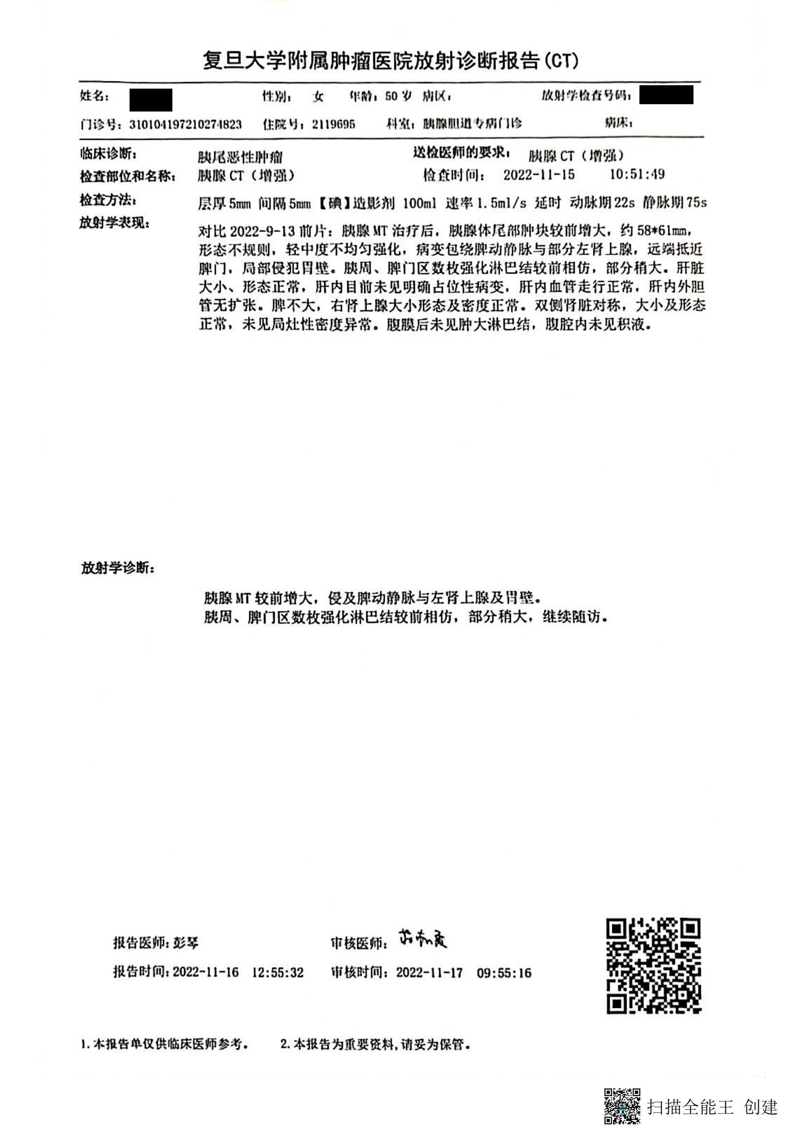
| 放射学表现:对比2022-9-13前片:胰腺MT治疗后,胰腺体尾部肿块较前增大,约58*61mm, |
| Radiological manifestations: Compared with the film got on September 13, 2022: After pancreatic MT treatment, the mass at the tail of the pancreas increased, about 58*61mm, |
| 形态不规则,轻中度不均匀强化,病变包绕脾动静脉与部分左肾上腺,远端抵近脾门,局部侵犯胃壁。 |
| Irregular in shape, mild to moderate uneven enhancement, the lesion surrounds the splenic artery and vein and partial left adrenal gland, the distal end is close to the splenic hilum, and partially invades the gastric wall. |
| 脾周、脾门区数枚强化淋巴结较前相仿,部分稍大.肝脏大小、形态正常.肝内目前未见明确占位性病变,肝内血管走行正常,肝内外胆管无扩张. |
| Several enhanced lymph nodes around the spleen and in the splenic hilum area are similar as before, some of them are slightly larger. The size and shape of the liver are normal. There is no definite space-occupying lesion in the liver at present, the blood vessels in the liver are normal, and the intrahepatic and extrahepatic bile ducts are not dilated |
| 脾不大,右肾上腺大小形态及密度正常.双侧肾脏对称,大小及形态正常,未见局灶性密度异常.腹膜后未见肿大淋巴结,腹腔内未见积液. |
| The spleen was not enlarged, and the size, shape, and density of the right adrenal gland were normal. The bilateral kidneys were symmetrical, with normal size and shape, and no focal density abnormalities. No enlarged lymph nodes were found in the retroperitoneum, and no fluid was found in the abdominal cavity. |
| 放射学诊断: |
| Radiological diagnosis: |
| 腴腺MT较前增大,侵及脾动静脉与左肾上腺及胃壁. |
| Pancreatic MT was larger than before and invaded splenic artery and vein, left adrenal gland and gastric wall |
| 脾周、脾门区数枚强化淋巴结较前相仿,部分稍大,继续随访. |
| Several enhanced lymph nodes in the perisplenic and splenic hilum area were similar as before, some were slightly larger, and the follow-up was needed. |
| 报告医师।彭琴审核医师: |
| Reporting Physician: Qin Peng Reviewing Physician: |
| 报告时间:2022-11-16 12:55:32 审核时同:2022-11-17 09:55:16 |
| Reporting time: Nov.. 16, 2022 12:55:32 Reviewing time: Nov. 17, 2022 09:55:16 |
| I.本报告单仅供临床医师参考.2.本报告为重要要资料,请妥为保管. |
| I. This report is only for reference of clinicians. 2. This report is important information, please keep it safe. |
| m扫描全能王创建 |
|
| 复旦大学附属肿瘤医院放射诊断报告(CT) |
| Radiological Diagnosis Report (CT) Of Cancer Hospital Affiliated To Fudan University |
| 姓名:***性别:女 年龄: 50 病区:放射学检查号码,11407881 |
| Name: *** Gender: Female Age: 50 Ward: Radiological Examination No., 11407881 |
| 门诊号:310101197210274823 住院号:2119695 科室:股腺胆道专病门诊病床। |
| Outpatient No.: 310101197210274823 Inpatient No.: 2119695 Department: Femoral gland and biliary tract disease outpatient Bed: |
| 临床诊断:胰尾恶性肿瘤送检医脚的娜,觑腺CT (增强) |
| Clinical diagnosis: Malignant tumor of the tail of the pancreas The Request of Examination Physican, pancreas CT (enhanced) |
| 检查部位和名称•胰腺CT (增强)检查时间,2023-01-2808:20:26 |
| Examination site and name: Pancreas CT (enhanced) Examination Time, Jan. 28, 2023 08:20:26 |
| 检查方法:层厚5mm间隔5mm【碘】造影剂100ml速率l.5ml/s延时 动脉期22s静脉期75s |
| Inspection method: layer thickness 5mm interval 5mm [iodine] contrast agent 100ml rate 1.5ml/s delay arterial phase 22s venous phase 75s |
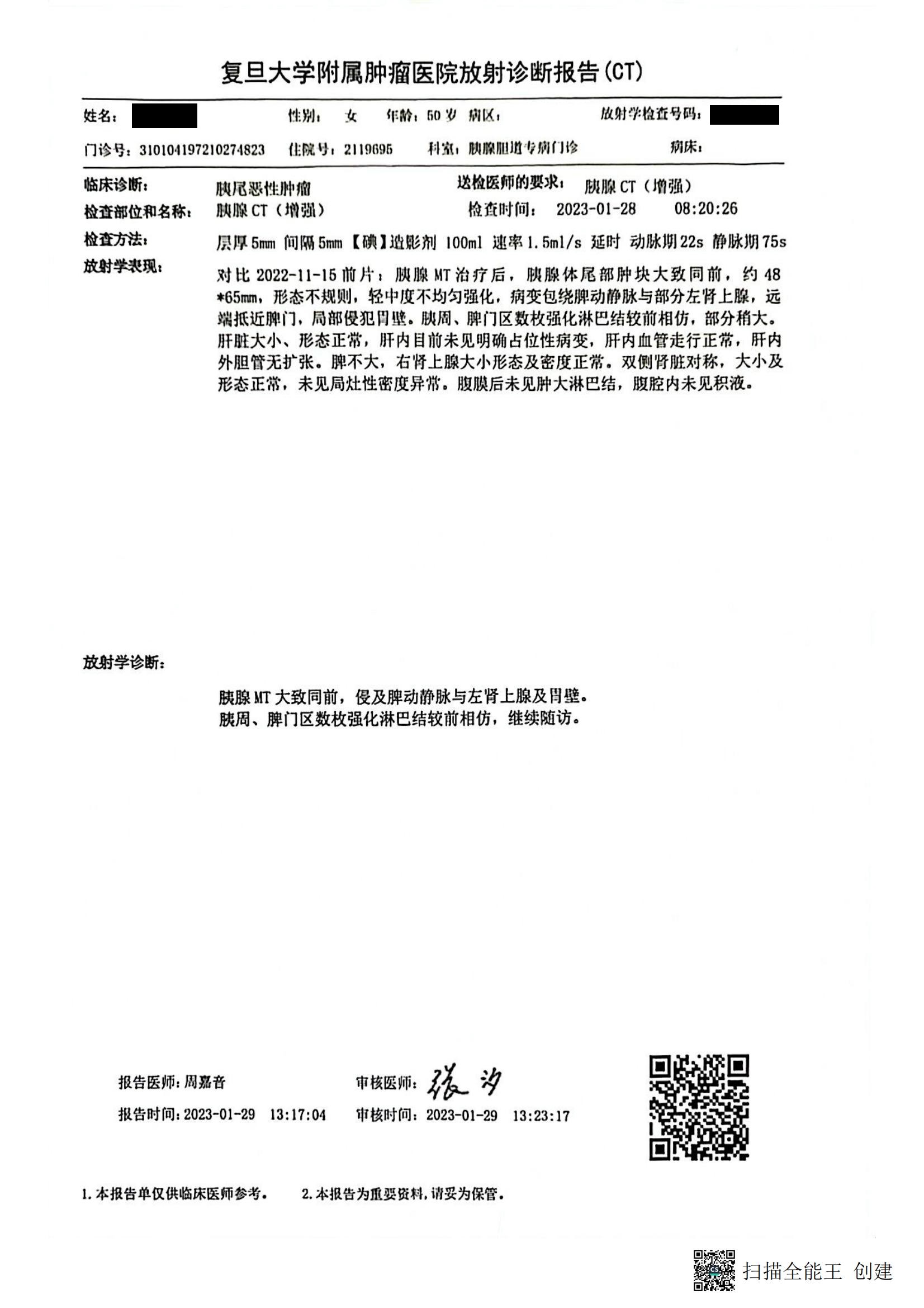
| 放射学表现,对比2022-11-15前片,腆腺MT治疗后,肤腺体尾部肿块大致同前,约48 |
| Radiological manifestations, compared with the previous film on November 15, 2022, After MT treatment of the pancreas, the mass in the tail of the pancreas is roughly the same as before,, about 48 |
| *65mm,形态不规则,轻中度不均匀强化,病变包绕脾动静脉与部分左肾上腺,远端抵近脾门,局部侵犯胃壁.胰周、脾门区数枚强化淋巴结较前相仿,部分梢大。 |
| *65mm, irregular shape, mild to moderate heterogeneous enhancement, the lesion surrounded the splenic artery and vein and some part of the left adrenal gland, the distal end approached the splenic hilum, and partially invaded the gastric wall. Several enhanced lymph nodes in the peripancreatic and splenic hilum area were similar as before, some of which are larger. |
| 肝脏大小、形态正常,肝内目前未见明确占位性病变,肝内血管走行正常,肝内外胆管无扩张.脾不大,右肾上腺大小形态及密度正常.双侧肾脏对称,大小及形态正常,未见局灶性密度异常,腹膜后未见肿大淋巴结,腹腔内未见积液. |
| The size and shape of the liver are normal, no clear space-occupying lesions are found in the liver, the blood vessels in the liver run normally, and the extrahepatic bile ducts are not dilated. The spleen is not enlarged, and the size, shape and density of the right adrenal gland are normal. , no focal density abnormalities, no enlarged lymph nodes in the retroperitoneum, no effusion in the abdominal cavity. |
| 放射学诊断; |
| Radiological diagnosis; |
| 胰腺MT大致同前,侵及脾动静脉与左肾上腺及胃壁。 |
| Pancreatic MT is roughly the same as before, invading the splenic artery and vein, left adrenal gland and gastric wall. |
| 胰周、脾门区数枚强化淋巴结较前相仿,继续随访。 |
| Several enhanced lymph nodes in the peripancreatic and splenic hilum area are similar as before, and the follow-up was needed. |
| 报告医师:周嘉音 |
| Reporting physician: Jiayin Zhou |
| 报告时间i 2023-01-29 13:17:04 |
| Reporting time: Jan. 29, 2023 13:17:04 |
| 审核医师: |
| Reviewing Physician: |
| 审核时间:2023-01-29 13:23:17 |
| Review time: Jan. 29, 2023 13:23:17 |
| 本报告单仅供临床医肺参考.2.本报告为重要资料,请妥为保管. |
| This report is only for clinical reference. 2. This report is important information, please keep it safe. |
| 扫描全能王创建 |
|


_05-scaled.jpg)
_01-scaled.jpg)
_02-scaled.jpg)
_03-scaled.jpg)
_04-scaled.jpg)
_05-1-scaled.jpg)
_06-scaled.jpg)
_07-scaled.jpg)
_08-scaled.jpg)


失业证明12.24_01-768x1086.jpg)
 | &pic=https://landeservice.oss-cn-shanghai.aliyuncs.com/wp-content/uploads/2023/03/诊断书徐(CN)_05-scaled.jpg&ralateUid=5788774445&language=zh_cn)
&summary=翻译中英文对照文本 seg上海中医药大学附属龙华医院出院小结Discharge Summary of Longhua Hospital Affiliated to Shanghai University of Traditional Chinese Medicine姓名:*** 科别:肿瘤七科 病区:二十六病区 床号:14住院号:0823817Name: *** Department: Oncology the 7th Department Ward No.: 26 Bed No.: 14 Hospital No.: 0823817更诊时间及后续治疗:门诊随访,定期复查,联系电话64385700-8163(医),8168 (护);我科来电显示:02133324000Time in service of hospital and follow-up treatment: outpatient follow-up, and regular review, Contact Number 64385700-8163 (medical), 8168…&url=https%3A%2F%2Fwww.landeservice.cn%2Farchives%2F33687.html&pics=https://landeservice.oss-cn-shanghai.aliyuncs.com/wp-content/uploads/2023/03/诊断书徐(CN)_05-scaled.jpg)